Additional facts

Recently, increasing reports suggest that up to 5% of patients with beta-thalassemias produce fetal hemoglobin (HbF), and use of hydroxyurea also has a tendency to increase the production of HbF, by as yet unexplained mechanisms.[citation needed]

Giving a happy ending to a poignant family tale and raising fresh hope of leveraging stem cell therapy, a group of doctors and specialists in Chennai and Coimbatore have registered the first successful treatment of thalassaemia in a child using a sibling's umbilical cord blood.

Read Users' Comments (0)

News

Life Expectancy Increased:

Due to improved treatments, many patients are living longer, but longer life expectancy has led to new problems. Thalassemia patients are now struggling with secondary conditions such as heart disease, hepatitis, liver cancer, osteoporosis, and fertility problems.

Thalassemia in Italy:

In Italy, the number of thalassemia major patients is estimated to be between 5000 and 8000.

Read Users' Comments (0)

Benefits of Thalassemia

Being a carrier of the disease may confer a degree of protection against malaria, and is quite common among people from Italian or Greek origin, and also in some African and Indian regions. This is probably by making the red blood cells more susceptible to the less lethal species Plasmodium vivax, simultaneously making the host RBC environment unsuitable for the merozoites of the lethal strain Plasmodium falciparum. This is believed to be a selective survival advantage for patients with the various thalassemia traits. In that respect it resembles another genetic disorder, sickle-cell disease.

Epidemiological evidence from Kenya suggests another reason: protection against severe anemia may be the advantage.

People diagnosed with heterozygous (carrier) Beta-Thalassemia have some protection against coronary heart disease.

Read Users' Comments (0)

Diet for Thalassemia

  • A well-balanced diet with adequate folic acid supply is a necessity. Foods with high iron content should be avoided, particularly meat because heme iron is especially well absorbed. Vitamin C assists absorption of dietary iron; patients should avoid co-ingesting vitamin C and iron-rich foods.
  • Alternatively, drinking tea with iron-rich foods helps chelate some of the iron before it is absorbed in the bowels.

Read Users' Comments (0)

Activity in Thalassemia

  • Many patients with thalassemia intermedia should be able to tolerate most daily activities. However, once the anemia worsens, exercise intolerance develops and may represent a warning sign indicating the need for initiation of blood transfusions.
  • Massive splenomegaly has been observed in severe cases and is a cause for limiting the patient's activity for fear of injury to the abdomen causing rupture of the spleen.
  • Regular transfusions decrease the size of the spleen in most instances, allowing splenectomy to be avoided whenever possible.

Read Users' Comments (0)

Consultations for Thalassemia

  • Patients in whom thalassemia is suspected should be seen and evaluated by a hematologist.
  • Consultation with a cardiologist is indicated to evaluate cardiac function and monitor potential complications due to the anemia, transfusion, or iron overload.
  • Consultation with an endocrinologist is also indicated for evaluation of possible involvement of various endocrine glands, which could result in diabetes mellitus, thyroid disorder, or growth retardation.
  • Patients should be seen by a gastroenterologist for diagnosis and management of liver complications.

Read Users' Comments (6)

Surgical Care in Thalassemia

  • Splenectomy is frequently recommended for patients who are no longer able to maintain an adequate Hb level. It is usually performed to restore the Hb steady state in patients who are not receiving blood transfusions and frequently succeeds in averting the need for regular transfusions.
  • Observations and case reports have shown that splenectomy in such patients may cause serious venous thrombotic events, ranging from deep vein thrombosis to pulmonary thrombotic lesions complicated by pulmonary hypertension. Several reports of serious thrombotic events such as transient ischemic attacks associated with hemiparesis and intracranial manifestations of Moyamoya syndrome were reported postsplenectomy in patients with thalassemia intermedia.For this reason, one should delay or reconsider such a procedure whenever possible. This is supported by the fact that many children who underwent splenectomy to avoid becoming transfusion dependent experienced only a transient effect, and most later required regular transfusions.
  • Placement of a central vascular access catheter in patients with severe disease is very helpful for blood transfusions, and laboratory work, especially when accessing a patient's peripheral veins becomes very difficult.
  • In the rare patient with large tumorlike masses that compress vital organs, surgical resection rather than radiation therapy is usually preferred.
  • Liver biopsy is indicated in the patient receiving chelation therapy for hemosiderosis to evaluate the degree of liver involvement and iron overload.

Read Users' Comments (1)comments

Medical Care in Thalassemia

The treatment of most cases of thalassemia intermedia involves close monitoring and observation.

  • Patients with satisfactory hemoglobin (Hb) levels are frequently monitored.
  • These patients usually require blood transfusions only on certain occasions such as the presence of intercurrent infections, hypersplenism, or other illnesses.
  • If patients can no longer maintain an Hb level of more than 6 g/dL, they are either started on a regimen of regular blood transfusions or a different option, such as splenectomy, should be tried. Patients with evidence of hypersplenism have a good chance to have their need for blood transfusion reduced or totally eliminated; this might last for months or years. Others may try to administer one of the drugs that may induce stress erythropoiesis and raise Hb levels. Hydroxyurea has been frequently used for this purpose. In separate studies on a large number of patients, a response rate exceeding 75% was reported after long-term therapy. However, this high rate of response was not confirmed by other studies until a study on a small number of patients with β thalassemia major and intermedia treated with hydroxyurea demonstrated a response rate of more than 82%.
    • The initial regimen includes transfusion of 10-15 mL of packed red blood cells (PRBC) every 4-5 weeks to keep the Hb level over 10 g/dL.
    • Blood transfusions should be leukocyte poor to avoid sensitization because such patients have the potential of becoming transfusion dependent in the future.
    • Patients should be checked and typed for minor blood groups to avoid further difficulties in providing appropriate blood for them in the future.
    • Identification of a small group of dedicated donors minimizes the risk of viral exposure and alloimmunization.
  • Iron status must be carefully monitored, and patients with iron overload should be treated with an aggressive chelation regimen as soon as indicated.
    • A popular chelation regimen includes administration of deferoxamine 5 days per week as a subcutaneous infusion over 8-12 hours. This regimen revolutionized the treatment of β thalassemia major in patients regularly receiving PRBC transfusions and resulted in longer survival and near-normal quality of life. An oral iron chelator, deferasirox (Exjade), has been in use in the United States for some time now. This agent has a long half-life and, for this reason, is orally administered once daily. Several studies have confirmed the long-term efficacy and safety of this agent. It is now much more popular and, due to the ease and convenience of administration and better compliance, is probably replacing deferoxamine
    • A similar dose is often administered at the time of blood transfusions to help bind the transfused iron (from hemolyzed RBCs).
  • Nutritional deficiencies should be addressed and treated.
    • A folic acid supplement should be administered.
    • Vitamin C supplementation has been effective in enhancing the efficiency of chelating iron from tissues.
  • Patients who have undergone a splenectomy should be placed on prophylactic antibiotics and be treated empirically for any signs of infection or fever while awaiting the results of blood cultures.
  • Appropriate vaccinations, including the polyvalent polysaccharide pneumococcal, the Haemophilus influenzae type b, and the quadrivalent meningococcal vaccines, should be administered to patients 1-2 weeks before splenectomy.
  • Patients with severe β thalassemia intermedia are prone to infection with Yersinia enterocolitica, similar to individuals with the severe forms of thalassemia major. For this reason, patients who develop fever without clear cause should receive appropriate treatment even if culture results are negative.
  • Young children should have their growth and development closely monitored; any deviation from normal should alert the physician to further investigate the need for blood transfusions.
  • Failure to thrive, exercise intolerance, bone deformities and fractures are all potential complications; the health care provider should always look for ways to prevent these complications or at least identify and treat them early with regular blood transfusions, which are frequently effective in reversing or preventing their progress.
  • In patients with severe thalassemia intermedia who require aggressive therapy to sustain life, bone marrow transplant, similar to that performed in patients with thalassemia major, is a reasonable alternative to transfusion and chelation if a matched sibling donor is available.
  • Many studies have shown that patients with thalassemia intermedia who are not on regular blood transfusion because of their milder symptoms nevertheless develop major complications related to their chronic anemia and ineffective erythropoiesis (IE). Considering the cost-benefit balance of regular treatment in patients with thalassemia major, most patients with thalassemia intermedia would apparently benefit from similar therapy to prevent the complications, rather than waiting to deal with such complications when they occur.

Read Users' Comments (0)

Folic acid (Folvite)

Required for DNA synthesis; therefore, patients with all conditions associated with rapid cellular turnover, such as hyperactive marrow in thalassemia, have greatly increased demand. Because use of folic acid in hemolytic anemias is extreme, deficiency states are fairly common in most of these patients. Patients who do not receive folic acid supplementation may develop megaloblastic anemia, increasing the severity of the original disease process.

Dosage:

Adult

1 mg PO qd

Pediatric

Administer as in adults

Interactions:

Increase in seizure frequency and a decrease in subtherapeutic levels of phenytoin reported when used concurrently

Contraindications:

Documented hypersensitivity; pernicious anemia; aplastic anemia

Precautions:

Pregnancy

A - Fetal risk not revealed in controlled studies in humans

Precautions

Pregnancy category C if dose exceeds RDA; benzyl alcohol may be contained in some products as a preservative (associated with a fatal gasping syndrome in premature infants); resistance to treatment may occur in patients with alcoholism and deficiencies of other vitamins.

Read Users' Comments (0)

Ascorbic acid (Cecon, Cevalin, Vita-C)

Vitamin C has been shown to enhance the function of deferoxamine by keeping iron in a form that can be chelated. When administered with deferoxamine, allows more iron to be removed.

Dosage:

Adult

100-200 mg/d PO during deferoxamine therapy

Pediatric

3 mg/kg/d PO with SC deferoxamine infusion

Interactions:

Decreases effects of warfarin and fluphenazine; increases aspirin levels

Contraindications:

Documented hypersensitivity

Precautions:

Pregnancy

A - Fetal risk not revealed in controlled studies in humans

Precautions

Use in patients with severe iron overload may induce a short-term deterioration with acute cardiac toxicity

Read Users' Comments (0)

Vitamins used in Thalassemia

These agents are compounds that are present in small amounts in food and are essential for normal metabolism, cell function, and healthy tissues.

Read Users' Comments (0)

Deferoxamine mesylate (Desferal)

Chelates iron from ferritin and hemosiderin but not from transferrin, cytochrome, or Hb. Helps prevent damage to liver and bone marrow from iron deposition.

Dosage:

Adult

1000 mg IV may be administered at a rate not to exceed 15 mg/kg/h; follow by a dose of 500 mg q4h for 2 doses; may administer additional IV infusion slowly over 24 h; not to exceed 6000 mg/d

Pediatric

20-40 mg/kg/d SC by infusion pump over 8-12 h 5 d/wk
With blood transfusions: 1-2 g IV slow infusion; not to exceed infusion rate of 15 mg/kg/h

Interactions:

Can cause loss of consciousness when administered with prochlorperazine

Contraindications:

Documented hypersensitivity; patients that do not have acute iron poisoning; severe renal disease and anuria (dose reduction after the loading dose should be considered in these circumstances)

Precautions:

Pregnancy

C - Fetal risk revealed in studies in animals but not established or not studied in humans; may use if benefits outweigh risk to fetus

Precautions

Tachycardia, hypotension, and shock may occur in patients receiving long-term therapy and could add to the cardiovascular collapse due to iron toxicity; GI adverse effects of the drug include abdominal discomfort, nausea, vomiting, and diarrhea, which may add to the symptoms of acute iron toxicity; flushing and fever are reported; increased susceptibility to Y enterocolitica infection.

Read Users' Comments (0)

Deferasirox (Exjade)

Tab for oral susp. Oral iron chelation agent demonstrated to reduce liver iron concentration in adults and children who receive repeated RBC transfusions. Binds iron with high affinity in a 2:1 ratio. Approved to treat chronic iron overload due to multiple blood transfusions. Treatment initiation recommended with evidence of chronic iron overload (ie, transfusion of about 100 mL/kg packed RBCs [about 20 U for 40-kg person] and serum ferritin level consistently >1000 mcg/L).

Dosage:

Adult

Initial: 20 mg/kg/d PO on empty stomach 30 min ac; as initial dose calculate dose to nearest whole tab
Maintenance: Adjust dose by 5- to 10-mg/kg/d increments q3-6mo according to serum ferritin level trends; not to exceed 30 mg/kg/d
Note: Dissolve tab completely in water, orange juice, or apple juice, then immediately drink susp; resuspend any remaining residue in small volume of liquid and swallow

Pediatric
<2>
>2 years: Administer as in adults

Interactions:

Data limited; do not take with aluminum-containing antacids

Contraindications:

Documented hypersensitivity

Precautions:

Pregnancy

B - Fetal risk not confirmed in studies in humans but has been shown in some studies in animals

Precautions

Common adverse effects include diarrhea, nausea, abdominal pain, headache, pyrexia, cough, and rash; may increase serum creatinine and hepatic enzyme levels; decrease dose with persistent elevation of serum creatinine level; may cause auditory and visual disturbances; slight decreases in serum copper and zinc levels may occur; dissolve tab completely in water, orange juice, or apple juice and drink resulting susp immediately (do not swallow tab whole, do not chew or crush); measure serum ferritin levels monthly and adjust dose every 3-6 mo based on serum ferritin trends.

Read Users' Comments (0)

Chelating agents

Chelating agents are an integral part of successful treatment of thalassemia. They remove excess iron deposits that are the main cause of long-term morbidity and mortality in this condition.

Read Users' Comments (0)

Acetaminophen (Feverall, Tylenol, Tempra)

Antipyretic effect through action on hypothalamic heat-regulating center. Although equal to aspirin in action, preferred because it has fewer adverse effects.

Dosage:

Adult

325-650 mg PO 30 min before transfusion

Pediatric

10-15 mg/kg/dose PO 30 min before transfusion

Interactions:

Rifampin can reduce analgesic effects of acetaminophen; coadministration with barbiturates, carbamazepine, hydantoins, and isoniazid may increase hepatotoxicity.

Contraindications:

Documented hypersensitivity

Precautions:

Pregnancy

B - Fetal risk not confirmed in studies in humans but has been shown in some studies in animals

Precautions

Hepatotoxicity possible in people with chronic alcoholism following various dose levels; severe or recurrent pain or high or continued fever may indicate a serious illness; APAP is contained in many OTC products, and combined use with these products may result in cumulative APAP doses exceeding recommended maximum dose.

Read Users' Comments (0)

Antipyretics, analgesic Medication for Thalassemia

These agents can help prevent febrile reactions in patients who are frequently transfused and thus may develop sensitization to blood products.

Read Users' Comments (1)comments

Vitamin E (Vita-Plus E Softgels, Vitec, Aquasol E)

MOA has been known for many years. In newborn or premature infants, in particular, deficiency has resulted in peculiar red blood cell morphology, leading to hemolysis; these changes are reversed by vitamin E. Peroxidation of membrane lipids by various oxidants, including iron-mediated oxygen radicals, is the main cause of this hemolysis and can be prevented by antioxidants such as vitamin E.

Dosage:

Adult

50-2000 IU/d PO

Pediatric

1 IU/kg/d PO

Interactions:

Mineral oil decreases absorption of vitamin E; vitamin E delays absorption of iron and increases effects of anticoagulants

Contraindications:

Documented hypersensitivity

Precautions:

Pregnancy

A - Fetal risk not revealed in controlled studies in humans

Precautions

Pregnancy category C if dose exceeds RDA; vitamin E may induce vitamin K deficiency; necrotizing enterocolitis may occur when large doses of vitamin E are administered.

Read Users' Comments (0)

Antioxidants used in Thalassemia

Vitamin E has been shown to help in decreasing iron-mediated toxic effects on cells by preventing or decreasing membrane-lipid peroxidation.

Read Users' Comments (0)

Trimethoprim-sulfamethoxazole (Bactrim, Septra, Cotrim)

By blocking tetrahydrofolic acid, selectively inhibits synthesis of nucleic acids and proteins by bacteria.

Dosage:

Adult

160 mg (trimethoprim)/800 mg (sulfamethoxazole) PO q12h (ie, one double-strength [DS] tab PO q12h)

Pediatric

<2 months: Do not administer
>2 months: 8-10 mg/kg/d (based on trimethoprim component) PO/IV divided q12h

Interactions:

May increase PT when used with warfarin (perform coagulation tests and adjust dose accordingly); coadministration with dapsone may increase blood levels of both drugs; coadministration of diuretics increases incidence of thrombocytopenia purpura in elderly patients; phenytoin levels may increase with coadministration; may potentiate effects of methotrexate in bone marrow depression; hypoglycemic response to sulfonylureas may increase with coadministration; may increase levels of zidovudine.

Contraindications:

Documented hypersensitivity; megaloblastic anemia due to folate deficiency; age <2>

Precautions:

Pregnancy

C - Fetal risk revealed in studies in animals but not established or not studied in humans; may use if benefits outweigh risk to fetus

Precautions

Do not use near term in pregnancy because of risk of kernicterus; discontinue at first appearance of skin rash or sign of adverse reaction; obtain CBC counts frequently; discontinue therapy if significant hematologic changes occur; goiter, diuresis, and hypoglycemia may occur with sulfonamides; prolonged IV infusions or high doses may cause bone marrow depression (if signs occur, administer 5-15 mg/d leucovorin); caution in folate deficiency (eg, people with long-term alcoholism, elderly people, those receiving anticonvulsant therapy, those with malabsorption syndrome); hemolysis may occur in individuals with G-6-PD deficiency; patients with AIDS may not tolerate or respond to therapy; caution in renal or hepatic impairment (perform urinalyses and renal function tests during therapy); administer fluids to prevent crystalluria and stone formation.

Read Users' Comments (1)comments

Penicillin V (Veetids)

DOC for prophylaxis in patients with thalassemia who have undergone a splenectomy (erythromycin used in patients allergic to penicillin); active against most microorganisms considered to be major pathogens in splenectomized patients (ie, streptococcal, pneumococcal, and some staphylococcal microorganisms) but not penicillinase-producing species. Prophylaxis provided for >3 y after splenectomy.

Dosage:

Adult

250-500 mg PO bid

Pediatric

<5 years: 125 mg/dose PO bid
>5 years: 250 mg/dose PO bid
Streptococcal infections: Administer above doses for >10 d
Prophylaxis: Treat for >3 y after splenectomy

Interactions:

Probenecid may increase effectiveness by decreasing clearance; tetracyclines are bacteriostatic, causing a decrease in the effectiveness of penicillins when administered concurrently.

Contraindications:

Documented hypersensitivity.

Precautions:

Pregnancy

C - Fetal risk revealed in studies in animals but not established or not studied in humans; may use if benefits outweigh risk to fetus

Precautions

Patients with asthma may have hypersensitivity; PO route usually not adequate for treatment of severe infections; treat for minimum of 10 d for streptococcal infections.

Read Users' Comments (0)

Gentamicin

An aminoglycoside. Effective against gram-negative aerobic microorganisms.

Dosage:

Adult

1-1.5 mg/kg IV q8h with normal renal function

Pediatric

6-7.5 mg/kg/d IV divided q8h

Interactions:

Coadministration with other aminoglycosides, cephalosporins, penicillins, and amphotericin B may increase nephrotoxicity; aminoglycosides enhance effects of neuromuscular blocking agents, thus prolonged respiratory depression may occur; coadministration with loop diuretics may increase auditory toxicity of aminoglycosides; possible irreversible hearing loss of varying degrees may occur (monitor regularly)

Contraindications:

Documented hypersensitivity; non–dialysis-dependent renal insufficiency

Precautions:

Pregnancy

C - Fetal risk revealed in studies in animals but not established or not studied in humans; may use if benefits outweigh risk to fetus

Precautions

Narrow therapeutic index (not intended for long-term therapy); caution in renal failure (patient not on dialysis), myasthenia gravis, hypocalcemia, and conditions that depress neuromuscular transmission; adjust dose in renal impairment.

Read Users' Comments (0)

Antimicrobial Agents

These agents are known to be effective against organisms that may cause infection in patients with iron overload who are also receiving deferoxamine therapy. Y enterocolitica infections are rare in healthy patients because the organism requires siderophores, which are present in patients with thalassemia but not in healthy patients. The appropriate therapy is a combination of trimethoprim-sulfamethoxazole and gentamicin. Patients who require splenectomy must receive prophylactic antibiotics to prevent fulminating sepsis, especially patients younger than 5 years.

Read Users' Comments (0)

Diphenhydramine hydrochloride (Benadryl, Benylin)

Elicits anticholinergic and sedative effects.

Dosage:

Adult

25-50 mg PO q6-8h prn; not to exceed 400 mg/d
10-50 mg IV/IM q6-8h prn; not to exceed 400 mg/d

Pediatric

Neonates and premature infants: Do not administer
Infants and children: 1 mg/kg/dose PO/IV q6h or 5 mg/kg/d PO/IV divided q6h

Interactions:

Potentiates effect of CNS depressants; because of alcohol content, do not administer syr dosage form to patient taking medications that can cause disulfiramlike reactions.

Contraindications:

Documented hypersensitivity; MAOIs

Precautions:

Pregnancy

C - Fetal risk revealed in studies in animals but not established or not studied in humans; may use if benefits outweigh risk to fetus

Precautions

May exacerbate angle closure glaucoma, hyperthyroidism, peptic ulcer, and urinary tract obstruction

Read Users' Comments (0)

Antihistamines

These agents prevent or ameliorate allergic reactions associated with transfusion of blood products.

Read Users' Comments (0)

Secondary Medication for Thalassemia

No specific medications are available for the treatment of thalassemia intermedia. Most patients with severe disease are prone to developing megaloblastic anemia due to folate deficiency for several reasons, including poor absorption, low dietary intake, and, most importantly, the extreme demand of the very active bone marrow for folic acid. For this reason, most patients benefit from a low dose of folate.

Many patients with thalassemia intermedia ultimately require regular blood transfusions, usually about every 3-5 weeks. Similar to patients with thalassemia major, patients with thalassemia intermedia who receive regular transfusions are usually premedicated with an antipyretic, such as acetaminophen, and an antihistamine, such as diphenhydramine, 30 minutes before transfusion to prevent both febrile and allergic reactions.

Patients with iron overload should be treated with chelation therapy (orally or parenterally). The drugs of choice at the present time are the oral agent deferasirox and deferoxamine administered subcutaneously by infusion pump 5 times per week. It can be administered while the patient sleeps. Low-dose vitamin C with each infusion of deferoxamine is beneficial in enhancing iron chelation. Combination therapy with more than one agent has proved to be effective in certain situations.

Patients with iron overload who develop fever of unknown origin may have Y enterocolitica infection. Treatment with gentamicin and oral trimethoprim-sulfamethoxazole should be initiated if no other cause for the fever is identified.

Hepatitis C virus (HCV) infection is the most common cause of hepatitis in patients with thalassemia. Because of the high risk of liver failure or even hepatocellular carcinoma in a liver already damaged by iron toxicity and frequent blood transfusions, HCV infection should be aggressively treated in these patients. Interferon alfa therapy has been effective in many children with HCV infection.

Other agents that may be of value in patients with thalassemia intermedia include vitamin E, which may prevent some of the toxic effects of the free radicals and other iron-related toxicity. Penicillin or one of its derivatives should be prophylactically administered for patients who have undergone a splenectomy. Some have also recommended a daily low dose of aspirin as prophylactic treatment in patients with thalassemia intermedia who underwent a splenectomy to prevent thrombotic events.

Read Users' Comments (0)

Medication

Medical therapy for beta thalassemia primarily involves iron chelation. Deferoxamine is the intravenously administered chelation agent currently approved for use in the United States. Deferasirox (Exjade) is an oral iron chelation drug also approved in the US in 2005.

The antioxidant indicaxanthin, found in beets, in a spectrophotometric study showed that indicaxanthin can reduce perferryl-Hb generated in solution from met-Hb and hydrogen peroxide, more effectively than either Trolox or Vitamin C. Collectively, results demonstrate that indicaxanthin can be incorporated into the redox machinery of β-thalassemic RBC and defend the cell from oxidation, possibly interfering with perferryl-Hb, a reactive intermediate in the hydroperoxide-dependent Hb degradation.

Read Users' Comments (0)

Exams and Tests Recommended

A physical exam may reveal a swollen (enlarged) spleen.

A blood sample will be taken and sent to a laboratory for examination.
Red blood cells will appear small and abnormally shaped when looked at under a microscope.
A complete blood count (CBC) reveals anemia.
A test called hemoglobin electrophoresis shows abnormal hemoglobin.

A test called mutational analysis can help detect alpha thalassemia that cannot be detected with hemoglobin electrophoresis.
Treatment

Treatment for thalassemia major often involves regular blood transfusions and folate supplements.

If you receive blood transfusions, you should not take iron supplements. Doing so can cause a high amount of iron to build up in the body, which can be harmful.

Persons who receive significant numbers of blood transfusions need a treatment called chelation therapy to remove iron from the body.

Bone marrow transplant may help treat the disease in some patients, especially children.
Outlook (Prognosis)

Severe thalassemia can cause early death due to heart failure a, usually between ages 20 and 30. Frequent blood transfusions with therapy to remove iron from the body helps improve the outcome.

Less severe forms of thalassemia usually do not result in a shorter life span.
Possible Complications

Untreated, thalassemia major leads to heart failure and liver problems, and makes a person more likely to develop infections.

Blood transfusions can help control some symptoms, but may result in too much iron which can damage the heart, liver, and endocrine system.

Read Users' Comments (0)

Preimplantation Genetic Diagnosis AND OUR FUTURE

By Joseph McPhee

(August 2003)

“Those who cannot remember the past are condemned to repeat it.”
— George Santayana

“In the realm of bioethics, the evils we face are intertwined with the goods we so keenly seek. Distinguishing good and bad thus intermixed is often extremely difficult.”
— Leon R. Kass MD, PhD
Chairman, The President’s Council on Bioethics

Since the announcement that the entire human DNA genome had been sequenced in June 2000, newspapers around the world have been rife with proclamations describing how this information is being used for the prevention and treatment of genetic disorders. Among the most promising and controversial claims is the ability of genetic technology to screen in vitro conceived embryos for the presence or absence of certain genes before implantation into the mother. However, accompanying these new technologies are a number of very old questions.

Lessons from the Past

Eugenics is not a new phenomenon. It is the term used to indicate the genetic improvment of the human race by controlled selective breeding [1]. Believers in eugenics seek to apply the techniques commonly used in animal husbandry and plant development to human beings. The eugenic movement was very popular throughout North America during the late nineteenth and early twentieth centuries [2]. The popularity of the eugenic movement can be attributed to a number of social and technological developments that coincided with the spread of the industrial revolution throughout the United States from approximately 1865 to 1890.

Science and technology made breathtaking strides in the latter half of the nineteenth century. So confident were people in the abilities of science to solve the problems of the world that in 1899 Charles Duell, head of the US patent office, suggested that his office be abolished, saying, “everything that can be discovered, has been discovered.” Clearly, Duell’s recommendation was arrogant and premature, but his statement reflects an attitude shared by many of his time. Life expectancies were rising thanks to the development of better public health practices. Incomes throughout the United States were rising due to the increased production afforded by industries at the time. Electricity was being made available throughout the country, increasing the overall quality of life for the newly affluent country.

However, in stride with these advances a number of disturbing developments were growing. Social upheavals throughout Europe led to increased unemployment among the working classes. Immigrants from Europe, particularly Eastern Europe, came to the U.S. seeking a better life. Although the U.S. needed immigrants and the cheap supply of unskilled labour they provided, the country was not prepared for the approximate one million people per year who were entering. Moreover, the jobs held by the immigrants were often the most dangerous, with high risk of illness or death. The rapid growth rate also led to many problems that persist during times of upheaval, such as alcoholism, prostitution, and an elevated crime rate. Darwin never really embraced eugenics as a way of dealing with societal problems, but others, observing problems associated with the rapid influx of immigrants, began to manipulate Darwin’s evolutionary theories as a way of rationalizing a controversial eugenic management method.

With the increased crime and poverty levels existing primarily amongst the recently arrived immigrants it was thought by many at the time that these problems existed because of a defect in these people [1]. According to them, it was society that ultimately paid the price for those who came to be known as “defectives” or “degenerates,” society deserved a solution. The irony was clearly lost on those at the time who looked to improve society but chose not to examine society itself. Thus, policies were developed that sought to limit the effect of inferior genes on American (read “white”) society [1]. These included immigration restrictions from certain countries, forced sterilization of inmates, marriage prohibitions, and racial segregation, the effects of which are still currently being felt.

Are Genetic Advances Leading Us Down the Same Road Again?

Although the science of eugenics was questionable in its interpretation of social reform and the mechanisms of heredity, it is important to note that at the time eugenic policies were developed, nothing was known about the nature of how traits were transferred from parent to offspring. Thus, one must consider the ignorance of the investigator who originally drew conclusions about controlling heritable traits in society.

Recent developments in reproductive genetic technology have raised concerns among some circles that the eugenic movement may be entering a renaissance [1]. However, this time around, rather than poorly defined diseases like “feeblemindedness,” we have clearly defined diseases with a known genetic component that can be identified, and in some cases treated. Orators of the new eugenics suggest another possibility, apart from identification and treatment, whereby diseases with a strong inherited genetic component can be eliminated entirely by preventing that gene from ever being passed on again.

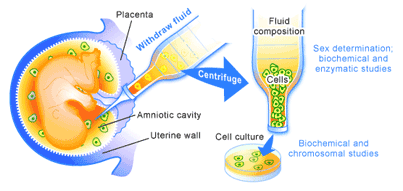
Pre-implantation genetic diagnosis (PGD) is an extension of previously existing prenatal screening technologies. Prenatal screening in an uncomplicated pregnancy usually involves ultrasound to examine fetal development and assess whether there is any abnormal development. In cases where these is a likelihood of abnormal development, as in women over the age of 35 or those with Down’s syndrome or spina bifida, chorionic villus sampling or amniocentesis may be performed. These techniques involve the removal of a sample of chorionic villus or amniotic fluid from a pregnant woman. A sample of which contain cells that have come from the developing embryo, which can be analyzed for the presence of abnormalities [5].

During the 1950’s and 1960’s, these techniques could only be used to determine the sex of the fetus or whether the cells contained sizeable chromosomal defects [5]. Thus, individuals at risk of transmitting X-linked diseases like Hemophilia could determine whether or not they carried a male child, or those at risk of pregnancies with a gross chromosomal defect like Down’s syndrome could examine the fetus for these types of genetic abnormalities. The difficulty of obtaining abortions at this time made the ability to look for these types of problems primarily a tool for preparing expecting parents for the likelihood of having a child with a chronic congenital disability.

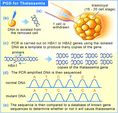
With advances in molecular biology in the 1970’s and early 1980’s, such as the development of polymerase chain reaction (PCR) and DNA sequencing, a much larger number of diseases were described at the genetic level. Soon to follow were tests for the presence of these diseases-associated genes, making prenatal screening a much more powerful tool [5]. These technological advances also coincided with the 1973 Supreme Court of the United States decision legalizing abortion on demand in that country.
Figure 1. Amniocentesis. Analyzing the biochemistry of the cells in the amniotic fluid.

In putting these two developments together, a moral conundrum was created. The combination made it possible to determine whether or not a child was healthy before it was born. Therefore, if a problem was found, that pregnancy could be terminated. For those individuals opposed to abortion, this was a disheartening development, since, in addition to the various social and economic reasons used to justify abortion, a new health-related reason was being created. Groups advocating the rights of people with disabilities foresaw the advancement of a Gattaca-like society, in which individuals without a desired genetic complement would be relegated to underclass status.

Gene Tests and Medical Science Today

Diseases that can be screened for today include cystic fibrosis, Tay-Sachs disease, Huntington’s disease, and a host of other devastating ailments [5]. Using PGD to screen against these diseases is relatively uncontroversial in the United States, because there is no regulation of fertility treatment. In the UK, the situation is somewhat different. A government body, the Human Fertilization and Embryology Authority (HFEA), was established in 1991 and monitors all labs providing fertility treatments and PGD [6]. It also licenses and monitors all embryo research.

In the UK, it is legal to screen in vitro fertilized (IVF) embryos for the presence of a disease gene and to implant only embryos that are healthy. However, improvements in screening have opened up a number of other applications for PGD. Take Thalassemia for instance. It is a genetic disorder that results in underproduction of the beta subunit of hemoglobin, resulting in a much lower ability to carry oxygen throughout the body. Treatment involves bimonthly blood transfusions, which unfortunately carry a side effect in the form of iron overload. This is treated by chelation therapy, but the disease is often fatal.

Figure 2. Prenatal Genetic Diagnosis for Thalassemia.


Treatment for the disease used to consist of bone marrow transplants, a treatment that is not very successful because the patient rarely survive the initial radiation treatment that allows the transplant to take place. More recently, umbilical cord blood has been shown to be a viable treatment for thalassemia [12]. By using PGD to screen embryos that are a perfect blood match for an affected individual (usually a sibling), the chances of finding a perfect match are greatly increased.

In the US, PGD was successfully used to screen embryos for both the absence of the Thalessemia disease gene and for a perfect tissue match for an older, diseased sibling [7]. The result was a healthy baby, with the umbilical cord blood transfusion giving his older sibling a 90% chance of survival. This marked the first case whereby a PGD was used not only to screen for the possibility of disease, but also for what could be termed a non-health related screening. Are we poised at the edge of a slippery slope? It is not a great leap to imagine that we are, and that screening for other “desirable” traits is close at hand. Since there is little regulation in private fertility clinics in the United States, it will require a great deal of political will to address this problem at this level.

Are we Already Sliding? The Case of the Hasmi Children

Indeed, the complexity of the issue is best illustrated by a recent series of court cases in Great Britain [8]. A couple from Leeds, Raj and Shahana Hasmi who have a son with thalassemia wanted another child. They realized that they were both carriers of the disease and wanted to ensure that their next child was free of the diseased thalassemia gene. To do this they wanted to use genetic technology to allow them to find an embryo that was free of the disease and that would be able to serve as a blood donor for their son. In this way, they hoped to have a healthy child and also harvest umbilical stem cells to cure their first son of his disease. If it worked in the US, they saw no reason why it couldn’t work in Britain.

Alarm bells started ringing at HFEA when the proposal for the technique was submitted for approval. The request was put on hold to allow the development of a policy toward the new technology. In December of 2001 policy was decided and the HFEA allowed the procedure to go ahead. However, one year later, following lobbying from the Comment on Reproductive Ethics (CORE), the British high court stepped in and ruled that the HFEA did not have the authority to allow PGD for the purposes of creating a perfectly matched donor [11]. The Hasmi’s were prevented from using the treatment. Not to be deterred, the Hasmi’s took their case to the Court of Appeal and again gained a ruling in their favour. In April of 2003, the HFEA, cautious after their previous decision had been overturned by the high court, ruled out widespread use of the technology but permitted the Hasmi’s to use the technology [10].

The period of time between the Hasmi’s application coming forward and the development of the current policy toward IVF and PGD, over 3 years, demonstrates the gap between the pace of technological advancements in genetics and the policies that will regulate it. Often the complexities of the science itself can mask the ethical questions that are raised by its application. In the case of the Hasmi’s, many questions are raised apart from the controversy associated with the use of the technology itself. What is the status of the embryo created? Is it a person with its own rights, or is it created merely as a means to an end? With the Hasmi’s intention to use umbilical cord blood there is no risk to the child that would result from the pregnancy. However, it is not difficult to envision a case in which embryonic stem cells would be the desired product. What would be permissible in this case? What if a kidney were required? A kidney transplant would entail some risk to the donor child, but failure to provide a perfectly matched kidney could result in the death of the older sibling. Even the fitness of the parents to make decisions on behalf of both of their children may be called into question. Many people foresee a future in which there will be two classes of children, the privileged who get to live, and others who are destined to be used for spare parts.

It is clear from this one case that there will be no easy answers to the questions that these developments have raised. In the UK, the back and forth battles among different regulatory agencies and judicial levels indicate that there will continue to be new developments in how these technologies will be used. It is heartening however, to note that the success of the HFEA in contributing to the development of strong policies on the use of PGD has prompted the development of similar agencies in other countries. While there is still no official agency dealing with these technologies in the United States, in November 2001 President Bush established the President’s Council on Bioethics [9]. Its first recommendations in July 2002 reflected the deep divisions that exist in the United States with respect to reproductive technology. In Canada, Health Canada has begun consultations to determine the structure of a regulatory agency to govern these technologies, but no unified policy has been released to date [10,11].

Society is a reflection of each member that it contains. It is very important when determining the applications of new technologies, to conduct debates in an open and honest way. Reproductive technologies present a number of very important and exciting opportunities for treating and preventing disease, but that excitement must be tempered by a full and frank discussion of what the consequences of their use may be. Just as those who once espoused eugenic principles could not foresee the negative outcomes of those policies, we must be wary of the possibility that decision we make now could seriously harm our society’s future. We must remain vigilant and engaged to ensure that this does not happen.

Additional Reading Material on Thalassemia:

1. Gould S.J. (1996). The Mismeasure of Man. New York: W. W. Norton Publishers. 444p.

2. Lynn R. (2001). Eugenics: A reassessment. Westport, CT: Praeger Publishers. 366p.

References
1. Lynn R. (2001). Eugenics: A reassessment. Westport, CT: Praeger Publishers. 366p.

2. Lombardo P. Eugenics Laws Restricting Immigration. Image Archive of the American Eugenics Movement.

3. Darwin C. (1859). The Origin of Species. London: J. Murray [1901]. 703p.

4. Darwin C. (1882). The Descent of Man. Amherst, NY: Prometheus Books [1998]. 698p.

5. The Genetic Drift Newsletter. 2001. Prental Diagnosis Vol.19 (Spring):

6. Human Fertilization & Embryology Authority

7. Belkin L. The Made to Order Saviour. New York Times Magazine. July 1, 2001.

8. BBC News. Family vow to have designer baby. Jan.10, 2003:

9. The President’s Council on Bioethics

10. Health Canada. (1999). Reproductive and Genetic Technologies Overview Paper

11. Health Canada, 2002. Press Release.

12. Goussetis E, Peristeri J, Kitra V, Kattamis A, Petropoulos D, Papassotiriou I, Graphakos S. (2000). Combined umbilical cord blood and bone marrow transplantation in the treatment of beta-thalassemia major. Pediatric Hematology & Oncology 17(4): 307-14.

Read Users' Comments (0)

Thalassemia Major

What Happens in Thalassemia Major?


Other Names for thalassemia major are:
Cooley's anemia
b-thalassemia major
Homozygous b-thalassemia
Homozygous thalassemia
Mediterranean anemia

How does thalassemia major first show itself?
During pregnancy the inherited thalassemia major does not affect the fetus. This is because the fetus has a special sort of hemoglobin, called "fetal hemoglobin" (HbF for short). Children and adults have a different hemoglobin called "adult hemoglobin" (HbA for shot). When a baby is born, most of its hemoglobin is still the fetal kind, but during the first 6 months of life it is gradually replaced with adult hemoglobin. The problem with thalassemia, is that the child cannot make adult hemoglobin. Therefore children with thalassemia major are well at birth, but usually become ill before they are 18 months old. They usually become quite anemic (their Hb level is usually less than 8 g/dl). So they become pale, do not grow as well as they should, and often have a big spleen.

The number of months that can pass before a thalassemic child becomes ill, can differ quite a lot from case to case. This is because thalassemia can be caused by several different defects in the hemoglobin genes. Some cause a complete absence of adult hemoglobin. While others allow a small amount of adult hemoglobin to be made. We call the first kind beta-zero thalassemia (b0-thalassemia for short). It is usually a very severe thalassemia, so the anemia begins early, at about 6-9 months of age. We call the second type beta-plus thalassemia, (b+-thalassemia for short). It is a little less severe. So affected children may stay well for a little longer. However, nearly all children with b-thalassemia major of any type become ill before 2 years of age, and need blood transfusions.

How do the tissues of a child with thalassemia manage to breathe, if there is no adult hemoglobin?
The child's body reacts to the shortage of adult hemoglobin by making some fetal hemoglobin, so most of the hemoglobin in your own blood is HbF. But your body is programmed to make fetal hemoglobin only in the fetus. It can only make a very small amount later on - not nearly enough to keep a child alive for long.


What happens if thalassemia major is not treated?
The anemia gets worse, the child stops growing altogether, and the spleen goes on getting bigger, so the tummy gets very big. the bone marrow, the tissue that forms the red blood cells, expands inside the bones, trying to make more and more red cells. But its efforts are useless. The red cells it makes do not contain enough hemoglobin, and simply die without ever getting of the bone marrow. However, the marrow's efforts to expand make the bones weak and alters their shape. The cheek bones and the bones of the forehead begin to bulge and the child's face gets a characteristics look, so that people can see from a distance that something is wrong. As time passes, the spleen, whose normal job is to destroy old red blood cells in the circulation, begins to destroy young red blood cells too, and finally also the white blood cells and the platelets. So in the end, the spleen makes the child's illness worse.


How do we treat thalassemia major?
Two different treatments are available at present, traditional treatment, and Bone-marrow Transplantation, which we describe in Chapter 4 of this Section. Here we will describe the traditional treatment. It consists of (1) Blood Transfusion, sometimes (2) Removing the Spleen, and (3) Desferal Treatment.

1. Blood Transufion:
To be precise, the treatment you need is not blood transfusion, but transfusion of red blood cells. You are only short of red blood cells, you make the other parts of the blood quite normally.
There are three reasons for blood transfusions.
(a) To correct your anemia. and make sure that your tissues get a normal amount of oxygen. This allows you to live and grow normally.
(b) To let your bone marrow rest, so that your bones can develop normally and you face looks normal.
(c) To slow down or prevent any increase in the size of your spleen, and to prevent hypersplenism.

2. Splenectomy:
When the spleen becomes too active and starts to destroy the red blood cell, transfusions become less and less effective. Then it may become necessary for a surgeon to take the spleen out. This operation is called "Splenectomy".

3. Desferal Treatment:
Every ml1 of red cells transfused brings 1mg2 of iron into the body. This iron can't be taken out of the blood because it is part of the hemoglobin, which is what your body needs. On its own, your body can only get rid of a tiny amount of iron, so if you have transufusions regularly, iron gradually accumulates in your body. It is stored in certain organs, especially the liver, the heart, and the "endocrine" glands (Figure 7). The iron behaves like a foreign body, and in the end would damage the organs where it is deposited. Fortunately, there are drugs that can pick up the iron, and carry it out of your body in your urine and feces. The only one that is used regularly at present is desferrioxamine, which is also called "Desferal". If you se Desferal regularly, you can keep the amount of iron in your body down to a safe level.
1ml stands for milliliter, one thousandth of a liter. You can see how much it is from the syringe you use for your Desferal infusions.
2One mg stands for one thousandth of a gram. Since one gram is one thousandth of a kilogram, you can see that one milligram is one millionth of a kilogram! It is not very much, but it adds up with time.

What happens when thalassemia is treated correctly?
For a well-treated patient thalassemia is quite different from the untreated disease. There is no anemia, growth is normal, and the face and the bones look nonmoral. However, there can be complications because of stored iron, or because of infections passed on in transfused blood. It is possible for viruses to get into your body with the blood and to make you ill.
In Particular, it is possible to get hepatitis, which is an infection of the liver, this way. It is also possible for the AIDS virus to be transmitted by transfusions, but this is very remote possibility, because all blood donors are first tested for AIDS. People who are found to be carriers, or who have been infected with the AIDS virus in the past are not allowed to give blood.

Read Users' Comments (0)

Splenectomy

Splenectomy (Removing the Spleen):
When the spleen becomes too active and starts to destroy the red blood cell, transfusions become lesser and less effective. Then it may become necessary to take the spleen out through surgery. This operation is called "Splenectomy".

Read Users' Comments (0)

Desferal Treatment

Blood transfusions bring extra iron into the body and if transfusions are regular, iron gradually accumulates in the body. It is stored in certain organs, especially the liver, the heart, and the endocrine glands. The iron behaves like a foreign body, and in the end would damage the organs where it is deposited. Fortunately, there are drugs that help the drainage of iron out of the body. The medication used very regularly is Desferrioxamine, more commonly called ‘Desferal’. Desferal keeps the amount of iron under a safe level in a Thalassemic’s body.

Read Users' Comments (0)

Bone-marrow Transplantation

A Thalassemic's bone marrow is not able to make a normal amount of red blood cells. If the malfunctioning bone marrow can be replaced with a normal bone marrow, this problem is solved.

At present, only young people with a fully compatible donor can have a bone-marrow transplant. A transplant in Pakistan (Zia-ud-Din Hospital, Karachi) or India (Apollo Hospital, Chennai) costs the equivalent of $13,000 to $14,000 (Pak Rs. 9-10 Lakh). A transplant in any other country may vary in cost.

Life Expectancy:

A difficult question perhaps, but Thalassemic’s and their families must be aware of this subject. The illness and its implications are changing almost from day to day, due to advances in treatment. With timely and correct treatment, Thalassemic’s live longer and healthier now. It is reasonable to think that people with Thalassemia, well treated from the beginning, may live as long as people without Thalassemia, mean an excellent life-expectancy.

Quality of life:

A chronic illness always causes some limitation of life, especially when it requires frequent and complex treatment, as Thalassemia does.

But still, the treatment should not be allowed to have a profound effect on a Thalassemic's life. In particular doctors and hospitals should make the effort to arrange out-patients visits for transfusions so that they interfere as little as possible with normal life. Treatment should not interrupt schooling or work. To manage this, some centers arrange transfusions on week-ends, others in late afternoon or at night.

Apart from a few cases, most Thalassemic’s lead a normal life. They go to school, take part in social activities and work, get engaged, and get married like everyone else. We are certain that, as time passes, the quality of life will steadily improve.

Read Users' Comments (0)

Blood Transfusion

Blood Transfusion:
Blood Transfusion is one of the most regularly practiced treatments for Thalassemia. To be precise, the treatment is not blood transfusion, but transfusion of red blood cells. These transfusions are necessary to provide the patient with a temporary supply of healthy red blood cells with normal hemoglobin capable of carrying the oxygen that the patient's body needs.

Today, most patients with a major form of Thalassemia receive red blood cell transfusions every two to three weeks. There are three reasons for blood transfusions.


a. To correct anemia and make sure that tissues get a normal amount of oxygen. This allows thalassemics to live and grow normally.
b. To let the bone marrow rest, so that the bones can develop normally and do not get deformed.
c. To slow down or prevent any increase in the size of spleen.

Read Users' Comments (0)

Treatment

Patients with thalassemia minor usually do not require any specific treatment. Treatment for patients with thalassemia major includes chronic blood transfusion therapy, iron chelation, splenectomy, and allogeneic hematopoietic transplantation.

Two different treatments are available at present, traditional treatment, and Bone-marrow Transplantation.

Read Users' Comments (0)

Genetic Prevalence

α and β thalassemia are often inherited in an autosomal recessive fashion although this is not always the case. Cases of dominantly inherited α and β thalassemias have been reported, the first of which was in an Irish family who had a two deletions of 4 and 11 bp in exon 3 interrupted by an insertion of 5 bp in the β-globin gene. For the autosomal recessive forms of the disease both parents must be carriers in order for a child to be affected. If both parents carry a hemoglobinopathy trait, there is a 25% chance with each pregnancy for an affected child. Genetic counseling and genetic testing is recommended for families that carry a thalassemia trait.

There are an estimated 60-80 million people in the world who carry the beta thalassemia trait alone. This is a very rough estimate and the actual number of thalassemia Major patients is unknown due to the prevalence of thalassemia in less developed countries in the Middle East and Asia where genetic screening resources are limited. Countries such as India, Pakistan and Iran are seeing a large increase of thalassemia patients due to lack of genetic counseling and screening. There is growing concern that thalassemia may become a very serious problem in the next 50 years, one that will burden the world's blood bank supplies and the health system in general. There are an estimated 1,000 people living with Thalassemia Major in the United States and an unknown number of carriers. Because of the prevalence of the disease in countries with little knowledge of thalassemia, access to proper treatment and diagnosis can be difficult.

As with other genetically acquired disorders, genetic counseling is recommended.

Read Users' Comments (0)

Prevalence

Generally, thalassemias are prevalent in populations that evolved in humid climates where malaria was endemic. It affects all races, as thalassemias protected these people from malaria due to the blood cells' easy degradation. Thalassemias are particularly associated with people of Mediterranean origin, Arabs, and Asians.[2] The Maldives has the highest incidence of Thalassemia in the world with a carrier rate of 18% of the population. The estimated prevalence is 16% in people from Cyprus, 1%[3] in Thailand, and 3-8% in populations from Bangladesh, China, India, Malaysia and Pakistan. There are also prevalences in descendants of people from Latin America and Mediterranean countries (e.g. Greece, Italy, Portugal, Spain, and others). A very low prevalence has been reported from people in Northern Europe (0.1%) and Africa (0.9%), with those in North Africa having the highest prevalence. Ancient Egyptians suffered from Thalassemia with as many as 40%[citation needed] of studied predynastic and dynastic mummies with the genetic defect. Today, it is particularly common in populations of indigenous ethnic minorities of Upper Egypt such as the Beja, Hadendoa, Saiddi and also peoples of the Delta, Red Sea Hill Region and especially amongst the Siwans.

Read Users' Comments (0)

Pathophysiology

The thalassemias are classified according to which chain of the hemoglobin molecule is affected. In α thalassemias, production of the α globin chain is affected, while in β thalassemia production of the β globin chain is affected.

Thalassemia produces a deficiency of α or β globin, unlike sickle-cell disease which produces a specific mutant form of β globin.

β globin chains are encoded by a single gene on chromosome 11; α globin chains are encoded by two closely linked genes on chromosome 16. Thus in a normal person with two copies of each chromosome, there are two loci encoding the β chain, and four loci encoding the α chain. Deletion of one of the α loci has a high prevalence in people of African or Asian descent, making them more likely to develop α thalassemias. β thalassemias are common in Africans, but also in Greeks and Italians.

Alpha (α) thalassemias:

The α thalassemias involve the genes HBA1 and HBA2, inherited in a Mendelian recessive fashion. It is also connected to the deletion of the 16p chromosome. α thalassemias result in decreased alpha-globin production, therefore fewer alpha-globin chains are produced, resulting in an excess of β chains in adults and excess γ chains in newborns. The excess β chains form unstable tetramers (called Hemoglobin H or HbH of 4 beta chains) which have abnormal oxygen dissociation curves.

Beta (β) thalassemias:

Beta thalassemias are due to mutations in the HBB gene on chromosome 11, also inherited in an autosomal-recessive fashion. The severity of the disease depends on the nature of the mutation. Mutations are characterized as (βo) if they prevent any formation of β chains (which is the most severe form of beta Thalassemia); they are characterized as (β+) if they allow some β chain formation to occur. In either case there is a relative excess of α chains, but these do not form tetramers: rather, they bind to the red blood cell membranes, producing membrane damage, and at high concentrations they form toxic aggregates.

Delta (δ) thalassemia:

As well as alpha and beta chains being present in hemoglobin about 3% of adult hemoglobin is made of alpha and delta chains. Just as with beta thalassemia, mutations can occur which affect the ability of this gene to produce delta chains.

In combination with other hemoglobinopathies:

Thalassemia can co-exist with other hemoglobinopathies. The most common of these are:
hemoglobin E/thalassemia: common in Cambodia, Thailand, and parts of India; clinically similar to β thalassemia major or thalassemia intermedia.
hemoglobin S/thalassemia, common in African and Mediterranean populations; clinically similar to sickle cell anemia, with the additional feature of splenomegaly
hemoglobin C/thalassemia: common in Mediterranean and African populations, hemoglobin C/βo thalassemia causes a moderately severe hemolytic anemia with splenomegaly; hemoglobin C/β+ thalassemia produces a milder disease.

Read Users' Comments (1)comments

Low Bone Mass in Thalassemia

Why is low bone mass an issue in thalassemia?
Having bones that grow and develop into strong,
healthy bones is important for everyone. Low bone
mass refers to weakness of the bones or bones that
are not as strong as they should be.
There are many factors that may determine why any
individual has low bone mass, including the genes
s/he inherited from parents, dietary patterns and the
amount of weight-bearing exercise the individual
engages in regularly. However, people with
thalassemia are also more prone to develop bone
mass difficulties due to factors specifically related to
thalassemia or its treatment, such as anemia,
overactive bone marrow, excess iron deposited in the
bones, reactions to deferoxamine and endocrinerelated
problems such as delayed puberty or
hypogonadism.
What is osteopenia?
Osteopenia occurs when a person has low bone mass,
but not so low as to be diagnosed with osteoporosis.
(Sometimes osteopenia is defined as “reduced bone
mass,” while osteoporosis is defined as “low bone
mass”).
The World Health Organization defines osteopenia as
occurring when a person’s Bone Mineral Density (BMD)
T-score is between -1 and -2.5. The WHO defines
osteoporosis as occurring when a person’s BMD Tscore
is lower than -2.5.
What is osteoporosis?
Osteoporosis is defined as a thinning of the bones,
with a reduction in bone mass due to a loss of
calcium and bone protein. In osteoporosis, bones
become porous and brittle, are unable to support
weight easily and fracture more readily.

What are problems associated with low bone mass?
A person with low bone mass, especially osteoporosis, is
more likely to experience fractures. Once fractured,
bones may take longer to heal or heal more poorly than
the bones of a person with normal bone mass.
Osteoporosis can affect a person’s posture, impair
physical activity and mobility and may create some
physical changes as the skeletal system becomes
increasingly affected.
Although any bone in the body can be affected by osteoporosis, the
bones most vulnerable to fracture tend to be in the hip, spine, wrist
and ribs.
How is low bone mass diagnosed?
Most people who have low bone mass are unaware of it; bone loss
may occur for a long time without any visible symptoms. As a result,
it is often undiagnosed until after a fracture occurs. Because it
occurs with such frequency in thalassemia, individuals with
thalassemia intermedia or thalassemia major should be checked
regularly by having a Bone Mineral Density (BMD) test on an annual
basis starting at around 8 years old. BMD is measured by a dual
energy x-ray absorptiometry test, commonly called a DEXA scan.
The BMD measurement will enable your doctor to determine your Tscore
or Z-score and to determine if you have osteopenia or
osteoporosis. The doctor should also check nutritional status and
vitamin levels (especially calcium and vitamin D).
What are T-scores and Z-scores?
A T-score measures a patient’s BMD against that of a normal, healthy
30-year-old. A score of “0” means a patient’s BMD is equal to that
of a normal, healthy 30-year-old. A score above 0 means the
patient’s BMD is greater than normal; a score below 0 means it is
lower than normal. As mentioned above, a score of -1 to -2.5
indicates osteopenia; a score lower than -2.5 indicates osteoporosis.
A Z-score measures BMD compared to a typical, healthy person
whose age is the same as the patient. Because low bone mass can
occur at a much younger age in thalassemia than in the general
population, a Z-score may provide a physician with information that
is more relevant in assessing bone mass in a person with thalassemia.
What can I do to prevent low bone mass?
Because thalassemia makes them predisposed to low bone mass,
people with thalassemia should take extra efforts to keep their bone
mass at healthy levels; some steps that can be taken include:
• An appropriate transfusion regimen, as determined with your
doctor. As anemia and overactive bone marrow are thought to
contribute to osteoporosis, keeping hemoglobin at an appropriate
level can decrease the risk of developing low bone mass.
• Maintaining adequate chelation therapy. Excess iron in the bones is
a factor in low bone mass, so rid the body of as much iron as
possible.
• Treatment of endocrine issues that may affect bone mass, such as
delayed puberty/hypogonadism. Some doctors may prescribe sex
hormones to treat the latter.
• Avoiding smoking.
• Regular exercise. Patients should first
discuss an appropriate exercise schedule with
their doctor that takes into account any
special needs before embarking on any
exercise routine. For the general population,
the National Osteoporosis Society
recommends that a person engage in three or
four 20-30 minute exercise sessions per week,
with the exercise focusing on weight-bearing
activities. For adults, some appropriate activities may include: brisk
walking, jogging, running, aerobics, step classes, dancing, circuit
training.
• Maintain a diet rich in calcium and vitamin D. This diet must also
take into account restrictions that patients may have in terms of
excess iron, heart issues, diabetes, or other factors; consulting a
nutritionist who understands your specific issues is advised.
Following are some foods and drinks that are good sources of calcium:
* Dairy products such as milk, yogurt and cheese (Note that skim
milk actually contains more calcium than regular milk)
* Dark green leafy vegetables such as broccoli, collard greens,
spinach, turnip greens, Brussels sprouts and bok choy
* Also tofu, okra, white beans, baked beans, rhubarb, peas, nuts,
whole wheat bread
Again, remember that selecting the right mix of calcium-rich foods
for your diet must also take into consideration other issues that may
affect your dietary choices.

• In addition to a diet rich in calcium and vitamin D, your doctor
may recommend taking calcium and/or vitamin D supplements.
Check with your doctor about the need for these supplements.
What can be done to treat low bone mass?
Following all of the above prevention measures is important in
treating low bone mass, to help insure that there is no further
decrease. In addition, some doctors may prescribe a drug in the
bisphosphonate family, such as Pamidronate or Etidronate. Some
research trials have indicated that IV administration of Zoledronic
acid may be beneficial to patients with thalassemia and
osteoporosis; other trials are investigating whether increasing zinc
intake may have a beneficial outcome for this patient population.
Future studies will prove helpful in assessing the value of these and
other options.

Read Users' Comments (0)

Carrier Detection

A screening policy exists in Cyprus to reduce the incidence of thalassemia, which since the program's implementation in the 1970s (which also includes pre-natal screening and abortion) has reduced the number of children born with the hereditary blood disease from 1 out of every 158 births to almost zero.
In Iran as a premarital screening, the man's red cell indices are checked first, if he has microcytosis (mean cell haemoglobin < 27 pg or mean red cell volume < 80 fl), the woman is tested. When both are microcytic their haemoglobin A2 concentrations are measured. If both have a concentration above 3.5% (diagnostic of thalassaemia trait) they are referred to the local designated health post for genetic counseling.

In 2008, in Spain, a baby was selectively implanted in order to be a cure for his brother's thalassemia. The child was born from an embryo screened to be free of the disease before implantation with In vitro fertilization. The baby's supply of immunocompatible cord blood was saved for transplantation to his brother. The transplantation was considered successful.

Read Users' Comments (0)

What is Thalassemia

Thalassemia (from θάλασσα, thalassa, sea + αἷμα, haima, blood; British spelling, "thalassaemia") is an inherited autosomal co-dominant blood disease. In thalassemia, the genetic defect results in reduced rate of synthesis of one of the globin chains that make up hemoglobin. Reduced synthesis of one of the globin chains can cause the formation of abnormal hemoglobin molecules, thus causing anemia, the characteristic presenting symptom of the thalassemias.

Thalassemia is a quantitative problem of too few globins synthesized, whereas sickle-cell anemia (a hemoglobinopathy) is a qualitative problem of synthesis of an incorrectly functioning globin. Thalassemias usually result in underproduction of normal globin proteins, often through mutations in regulatory genes. Hemoglobinopathies imply structural abnormalities in the globin proteins themselves.[1] The two conditions may overlap, however, since some conditions which cause abnormalities in globin proteins (hemoglobinopathy) also affect their production (thalassemia). Thus, some thalassemias are hemoglobinopathies, but most are not. Either or both of these conditions may cause anemia.

The disease is particularly prevalent among Mediterranean people, and this geographical association was responsible for its naming: Thalassa (θάλασσα) is Greek for the sea, Haema (αἷμα) is Greek for blood. In Europe, the highest concentrations of the disease are found in Greece and in parts of Italy, in particular, Southern Italy and the lower Po valley. The major Mediterranean islands (except the Balearics) such as Sicily, Sardinia, Malta, Corsica, Cyprus and Crete are heavily affected in particular. Other Mediterranean people, as well as those in the vicinity of the Mediterranean, also have high rates of thalassemia, including Middle Easterners and North Africans. Far from the Mediterranean, South Asians are also affected, with the world's highest concentration of carriers (18% of the population) being in the Maldives.

Source: Wikipedia

Read Users' Comments (0)

Smowtion Media